Here is a video of how the female human eggs look like under a microscope.
Human Eggs known as Ovum cannot be seen with naked eyes because they are very small.
According to the National Library of Medicine, they have an average diameter of 0.1mm.
They are about the size of a very small grain of sand.

Illustration of how a 0.1mm sand looks like. Source: https://www.mdpi.com/
Here is another microscopic view of the ovum.
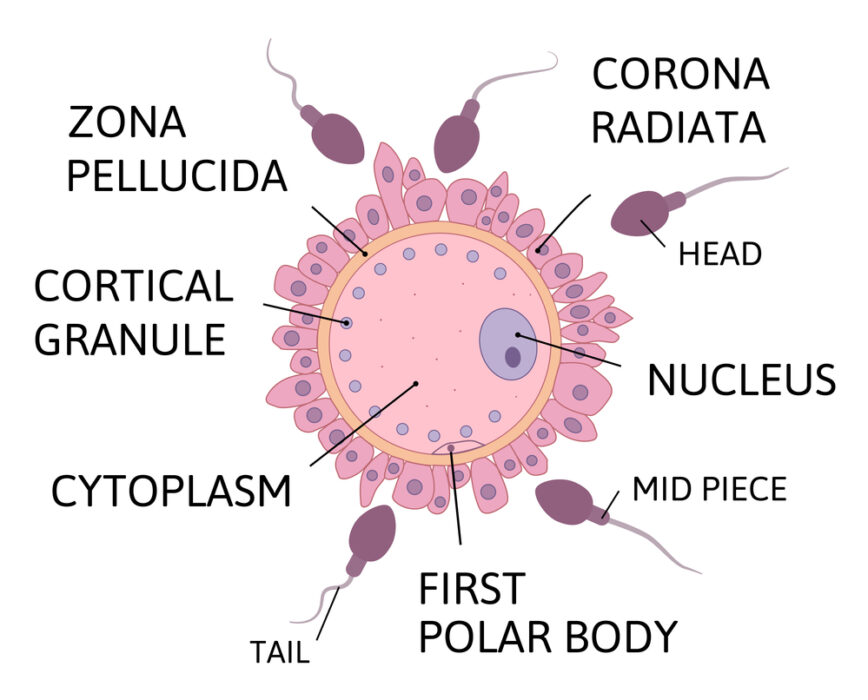
Illustrations of the Human Eggs (Ovums)
For a better view of what you saw under the microscope, see the image below.


Source: depositphotos.com
Recommended: Ovulation Calculator Based on Last 3 Periods
Layers of the Human Eggs

Photo Credit: Depositphotos.com
The Role of Microscopy
To truly grasp the appearance of female human eggs, advanced microscopes are essential. Several types of microscopes are used for this purpose, including:
- Optical Microscope (Light Microscope): Optical microscopes use visible light to magnify specimens. They are commonly employed to observe human eggs and provide clear images of their structure.
- Scanning Electron Microscope (SEM): SEMs are powerful instruments that use electron beams to create detailed 3D images of objects at a nanoscale level. SEMs have been used to study the ultrastructure of human eggs, revealing intricate surface details.
- Transmission Electron Microscope (TEM): TEMs are another type of electron microscope that provides high-resolution, cross-sectional images of specimens. They have been used for in-depth studies of the internal structures of human eggs.
